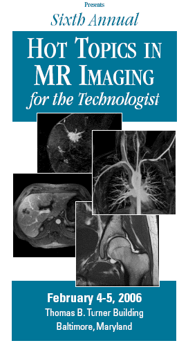
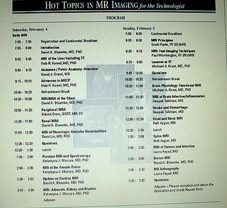
The Sixth Annual Hot Topics in MR Imaging for the Technologist,February 4-5,2006 Johns Hopkins Medical School, Baltimore,Maryland,USA
by Christopher A. Jankowski R.T.(R)(MR)ARRT
Hello my fellow Technologists,
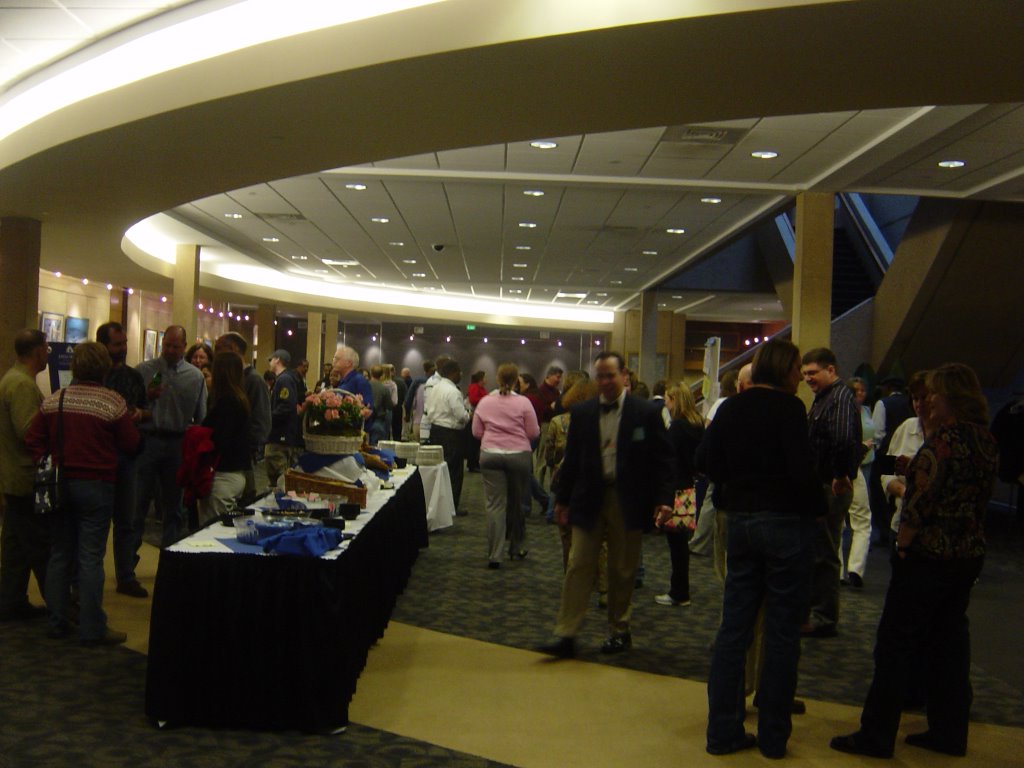
I had an opportunity to attend the seminar "The Sixth Annual Hot Topics in MR Imaging for the Technologist" taking place at Johns Hopkins Medical School in Baltimore,Maryland. I was the only one from Minnesota who joined more than 100 Technologists from all the States for the meeting.

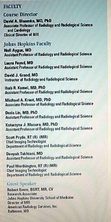
All lectures were presented by Johns Hopkins Radiologists alone with two Chiefs MR Radiologic Technologists .(see the window with JH Faculty)
We had a pleasure to meet the best of the best. Faculty of Johns Hopkins Radiology and Radiological Science kept us busy to the last minute of the each lecture.
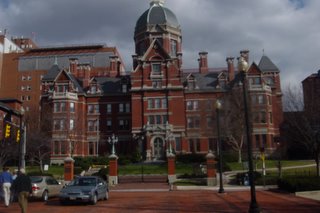
The first day started early around 06:00 am ,the hotel shuttle took us to the seminar site The Dr. T Turner Building. Dr .David A. Bluemke MD,PhD course director did the introduction ,right after
the breakfast on Saturday morning.
Dr.Kamel,MD,PHD ,a radiologist was the first speaker who talked about MRI of the liver including 3T. He reviewed the gross anatomy of the liver and outlined the physiology,techniques of imaging the abdominal region of the human body.
He spent a great amount of time on the arterial,portal and venous phase of imaging a liver,CT vs MRI detection and characterization,MR Imaging techniques with T1,T2 contrast enhancement sequences ,advantages and disadvantages with 1.5T and 3T.
Each of the speakers presented the Johns Hopkins MR protocols appropriate to the lecture.
Next was Dr.David J. Grand MD was talking about Abdomen/Pelvis Anatomy and
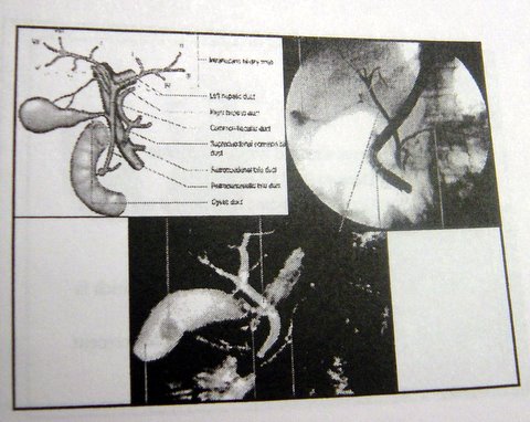
general imaging areas. Than we moved to the Advanced Imaging MRCP presented by Dr.IhabR.Kamel,MD,PhD.We had a chance to review the beautiful MR images of pancreative region of the human body.Just to let you know some centers in US gives the patients a pineapple juice to drink before the MRCP
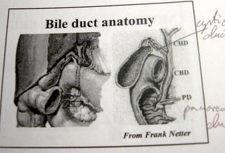
for better visualization of the bile ducts.
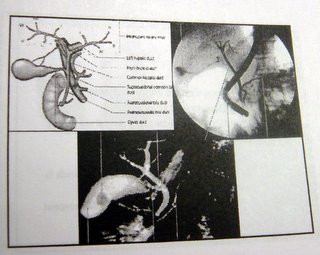
Interesting idea. I think we have to explore that possibility . We could see a lot of images with a great variety of pathology in that region.
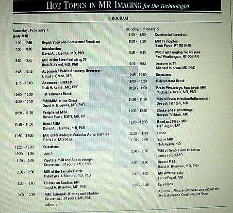
Here is the list of the two days of intensive lectures..all Technologist could enjoy it.
Than Dr. D.Bluemke MD,PhD presented slides
of the MRI/MRA of the Chest.
He went through the variety of MR techniques,protocols and imaging.
Our guest speaker was Robert Evans, from JH University School of Medicine. Very interesting gentleman, full of information and full of helpful hints how to avoid the pitfalls of some exams. Be sure to over lap you ROI coverage for some advance vascular exams. Robert was very positive and full of good humor what helped us to stay alert until the next coffee break.

He also showed us a very intriguing sequence called FBI Fresh Blood Imaging,made by Toshiba.
He spoke about non contrast 2DTOF techniques also talked about key technical consideration for carotids,run-offs,renal s,different approaches to the bolus capture.
Next we focused more on Renal MRA imaging,presented by Dr.David A. Blumke MD,PhD. He spoke with a great deal of details about renals MRA . He pointed out how important is to have the correct settings for slice thickness and the location of the slabs you will be able to image the affected part of anatomy and how valuable are the scout images. It is very important to position the center “below” the renal arteries to avoid vessel cut-off. Please remember the 3D time equals Te x Np x NEX x slices. In some instances is very important to create a 3D model of the patients renal arteries to help the radiologist to diagnose renal artery stenosis.
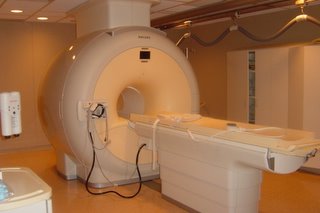
Johns Hopkins has 10 MR scanners 1.5 and 3T

,sharing patients between GE,Siemens and Phillips vendors.
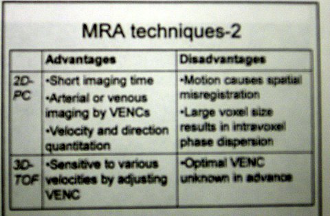
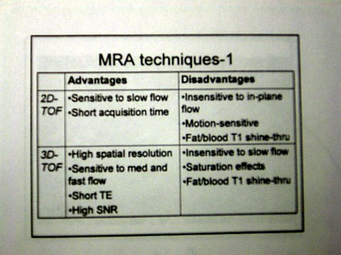
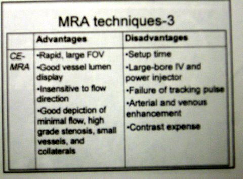
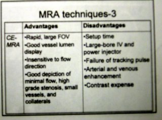
Our next lecturer was Dr.Doris Lin,MD,PHD she spoke about the MRI of Neurologic Vascular Abnormalities .She reviewed the applications of MR angiography in the evaluation of the brain and it's arteries. She presented advantages and disadvantages of MRA techniques
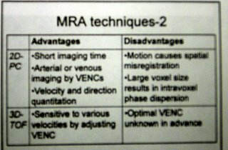
2D-PC,3D-TOF,CE-MRA

The next radiologist was Dr. Katarzyna J. Macura ,MD,PhD. She reviewed the standard MR imaging and spectroscopy for the evaluation of the prostate. Dr. Macura explained the prostate zonal anatomy,imaging techniques and the images of the prostate obtained by using the prostate coil. The main role of MRI in that exam are: presentation of extra capsular involvement,seminal vesicle involvement,nodal metastases and bone marrow metastases.Dr.Macura said they use the US gel to opacify the rectum and vagina for better assessment of those organs,especially when prolapse is to be
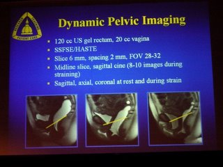
diagnosed,patients need to empty rectum for a complete evaluation when rectum without contrast/gel it cannot be well assessed we only use gel for dynamic pelvic floor MRI rarely for other indications,she said.She gave us very interesting view of DCE-MRI,DWI and spectroscopy of the prostate.
Than Dr. Macura moved her lecture to review the anatomy,pathology and MR imaging techniques for the female pelvis. The indications for the female pelvis imaging are benign and malignant tumors,abnormal pregnancy and pelvic floor imaging. She presented a variety of coils which we could use. She recognized the important of specific pulse sequences we should use,as follows: both T1,T2 for pathology detection and characterization ,T1 weighted imaging conventional SE,breath-hold GRE(spoiled GRASS or FLASH),SE higher SNR,GRE -multiple sections in a single breath hold when more extensive imaging needed. We need to remember to have patient 6 hours npo for those exams. Also we have to remember about the scanning plane of the interested region of the anatomy.
Many of the pelvis abnormalities were presented as :congenital uterine anomalies,septate bicornuate uterus, leiomyomas, submucosal fibroid, pre and post embolization, adenomyosis and more.
Last two lectures were about the Update on Cardiac MRI by David A. Bluemke,MD,PhD and MRI :Adrenals,Kidney and Bladder by Katarzyna J.Macura,MD,PhD. Dr.Bluemke talked about the essentials of Cardiac MRI,pulse sequences for cardiac MRI,than Dr. Macura focused on the anatomy,pathology and MR imaging techniques for the adrenal glends,kidney and urinary bladder.
That was only a very short summary of the seminar ,
our heads were spinning and our k-spaces filling up to the maximum...Sunday was even more fun...You can review more pictures from the seminar on that blogger below.

Next morning we were on the bus early again... hungry for more MRI advanture .(more to come in the next few days)
The next day our first speaker was Scott Pryde,R.T.(R)(MR) Chief Radiologic Technologist ,he talked about MRI Principles like signal formation,number of different techniques available today: 3d_volume acquisition,diffusion tensor imaging,arterialspin labeling,fat suppesion,parrallel imaging and multi-channel systems. Next Paul Worthington R.T.(R)(MR) Chief Radiologic Technologist discussed aspects of imaging resolution as they pertain to fast imaging techniques and methods and applications of fast imaging.
Dr.Michael A. Kraut,MD,PhD Lessons at 3T.He pointed out to the cost of the magnet. How we can use the field-strength dependent phenomena,how to take advantage of those phenomena,what to do with doubled SNR.
The images I could have seen were just incredible but for example the spine work was as good as on 1.5 T not particularly better.
Dr.Deepak Takhtani,MD presented
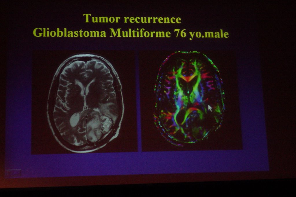
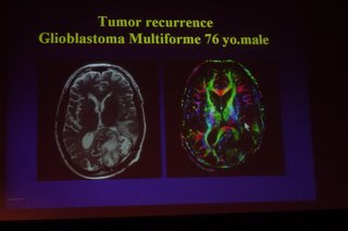
images of the Brain Infection/Inflammation and Stroke and Hemorrhage.
Dr.Nafi Aygun,MD Head and Neck MRI.Please review the pictures below.
Dr.David A. Bluemke,MD,PhD Br discussed optimal MRI methods for breast pathology. He did presentation on “accepted”indications for the breast MR,protocols and importance of using the phased array breast coil.
The most useful sequences are coronal, sagittal scout T1, sagittal FSE T2,3D”dynamic imaging”with a ZIPx2 and ZIP 512 options.Also is very important to use a light compression to reduce the motion. Later he presented a lot of MR images with a variety of different pathology.
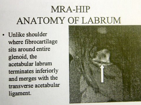
Dr.Laura Fayad,MD discussed the utility of direct and indirect MR arthography.Direct MRA advantages would be ,with direct injection,:distension of the joint capsule,after the injection contrast outlines intra-articular anatomy and leaks into abnormalities. Direct MRA disadvantages would be :invasive ,requires scheduling 2 modalities,risk of needle placement,it is more expensive. She presented the images of an elbow,shoulder,

hip,wrist,knee and the ankle.
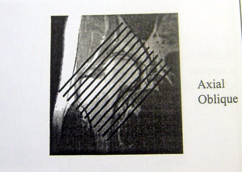
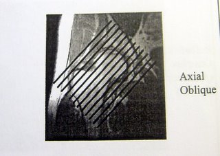
She showed us very interesting view axial oblique of the hip post injection .The hip labrium is very well presented in that view. All JH radiologists are using that image for reading with the confidence.
Indications for Indirect MRA are same as for direct MRA but provides additional/different information. The delay time would be :wrist,elbow,ankle 5-10 min,shoulder,hip 15-20 min and the knee more than 30 min. You also have to remember if you suspect tense joint effusion,increase the time.
Thank you all for your patience with me,if you got that far I am hoping you could learn or refresh your knowledge about MRI today.
I wanted to present all information from the seminar to all Technologists as much as I could. Please review more images from the seminar which I included at lower part of the blogger.
I created that web site/blogger for everybody who is interested in expending his/her horizon in the Art of MRI imaging.
Please go to my advertisement section to support the Free Blogger access and my links where you can find a lot of additional information about MR and more..
I also wanted to thank the St.Paul Radiology Administration for ongoing support in the Technologists education.
I wish everybody safe scanning and have a great days to come...
I am a Staff MR Technologist at St. Paul Radiology in St. Paul, Minn. I can be reached at christopheraj@yahoo.com or christopheraj@poczta.wprost.pl.